From Metal Fractures to Insects in Your Tea, Microscopy Reveals What Our Eyes Cannot See
By Carl Leblanc, Chemist, M.Sc., Metallurgist
For materials and chemistry experts, the microscope is an indispensable tool. Among other things, it helps us to understand failure mechanisms, observe material characteristics, trace the source of contamination, and determine both the nature and failure mode of components. In fact, the microscope is usually the first tool used by an expert in order to establish a diagnosis and understand a failure mode.
Different types of microscopes can be utilized. From the binocular microscope, which will provide the expert with a tridimensional view, to an electron microscope, which will use an electron beam instead of light. The electron beam offers a higher magnification along with elemental analysis capabilities, making it possible to identify the chemical elements found in the samples under study.
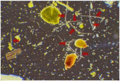
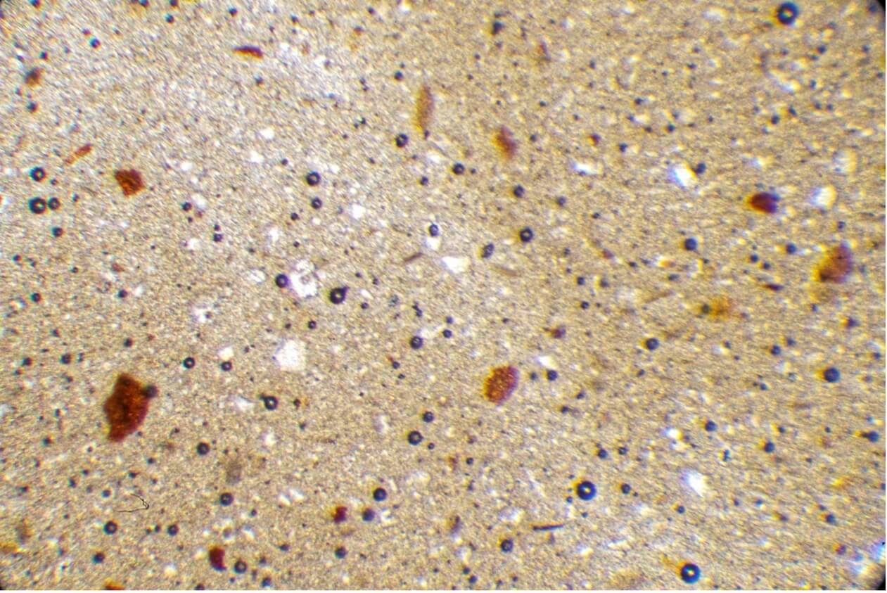
What can an optical microscope reveal about everyday products? Let’s see what can be found in tea (Figure 1) and peanut butter (Figure 2). A microscope examination of these two products enables the observation of structures that are invisible to the naked eye. Indeed, tea contains a significant amount of insect parts whereas peanut butter will show a noteworthy concentration of inorganic particles (soil and/or sand grains) resulting from the product’s transformation.

Figure 1 – Microscopic examination of green tea.
The structures with a tubular morphology (indicated by the arrows) are insect parts (setae) that were present during the tea processing.

Figure 2 – Microscopic observation of peanut butter.
The white crystalline structures visible throughout the image are soil or sand particles.
Component Fracture

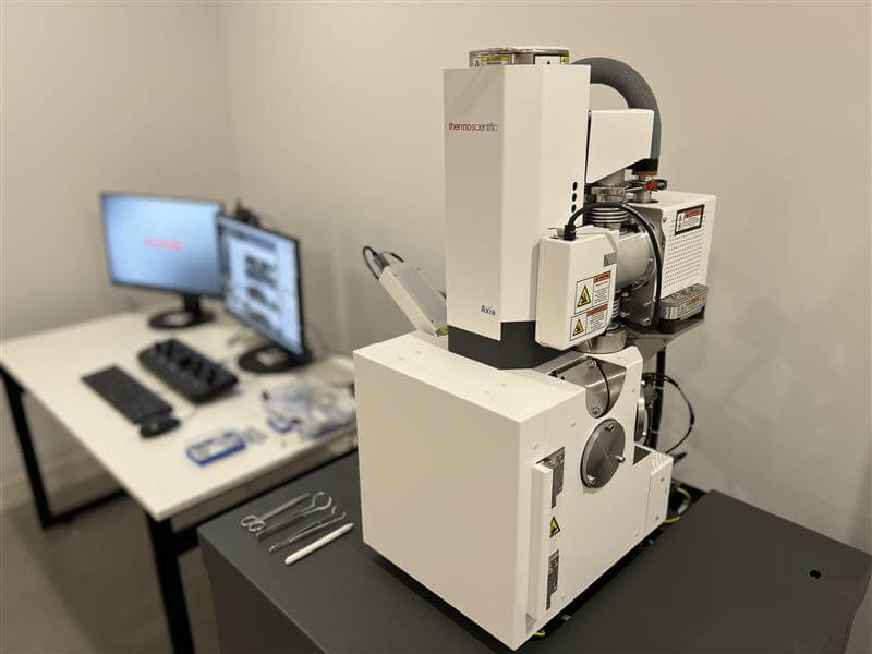
Figure 3 – Scanning Electron Microscope (SEM)
A fracture examination with a binocular microscope, with low magnification, allows an expert to analyze the failure mode and determine if it was sudden or progressive.
Moreover, a scanning electron microscope (SEM), offering a higher magnification, can provide us with more precise details about the nature of the failure as well as the possibility of a manufacturing defect. Coupled with energy-dispersive spectroscopy (EDS), the analysis will be able to let us know if corrosive elements, such as chlorine, could have contributed to the failure.
Soot Contamination
The presence of black deposits on interior surfaces of a building may indicate soot contamination. By using an appropriate sampling method, an optical microscope can confirm the presence of soot and analyze the morphology to determine the source. By doing this, we can determine whether the soot originated from a neighbouring source or from an external fire (such as a grass fire or a forest fire), or from excessive use of low-quality candles. This type of analysis is particularly useful to claims adjusters, as it allows them to precisely delineate the areas contaminated in the aftermath of a fire.
Water Infiltration During Merchandise Transportation
In a globalized economy, water infiltration can cause major property damage. Once again, an SEM, combined with an EDS, can be used to analyze water-related damage and determine its origin. For instance, by analyzing water-contaminated surface samples, it would be possible to determine whether it occurred at sea or during rail transport.
Component or Product Contamination
Chemical analysis of a contaminant can be carried out using a microscope that relies on infrared light rather than visible light. This technique makes it possible to identify the chemical compounds present in a contaminant and thereby determine its origin. It is particularly relevant in cases involving failures of CPVC fire sprinkler pipes, which may have been caused by the presence of internal or external contaminants, by contact with an incompatible sealant, or by direct contact with an electrical cable.
Car Damage
By combining the results of both infrared and electron microscope analyses, it is possible to determine whether damage observed on a vehicle resulted from contact with another vehicle (through paint transfer analysis) or with a fixed structure, such as a building. This type of expertise also allows us to confirm if the impact is the result of an attempted fraud or a genuine accident.
Glass and Sealed Glass Units
Finally, the combined use of a polarized light microscope, an infrared microscope, and an SEM, allows experts to identify or confirm the cause of glass breakage, whether we’re talking about the windows of a building or other glass objects like an aquarium. These techniques can also help determine the origin of chemical fogging inside a sealed glass unit, or the degradation of its internal energy-efficient coating. In addition, they can be used to pinpoint the exact source of scratches on glass panels by analyzing the microscopic deposits found within the scratches themselves.
Conclusion
The various microscopy techniques—and their combined use—are powerful tools that support experts in determining the exact origin and cause of the observed damage.
Do you have special requests for microscopic observations that spark your curiosity? Don’t hesitate to reach out to CEP Forensic!
